A simple method for making a gill section

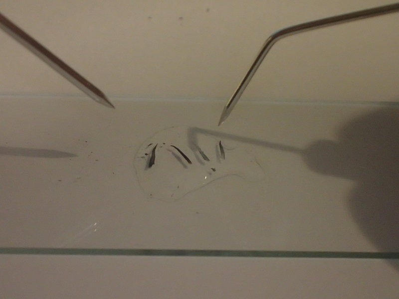
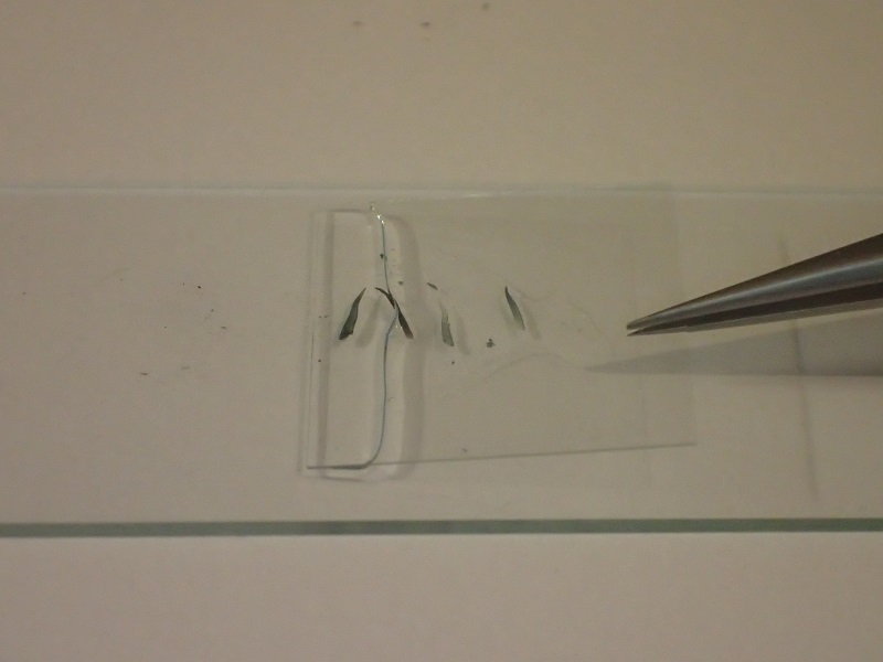
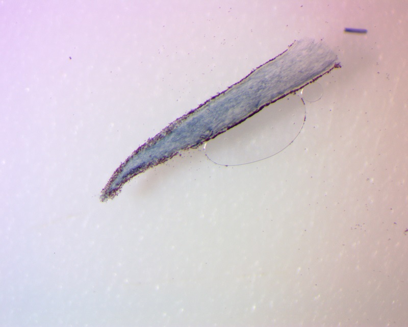
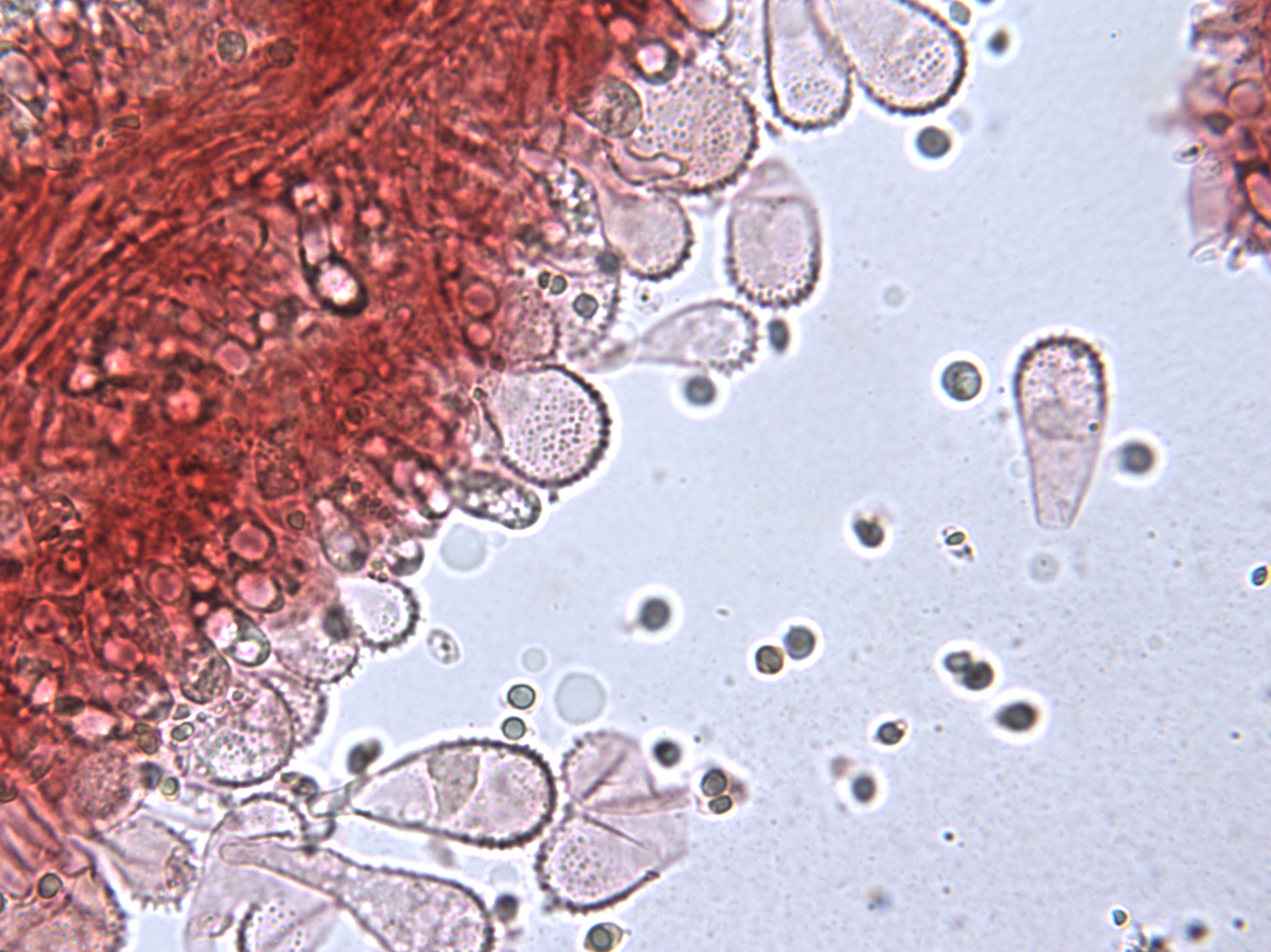
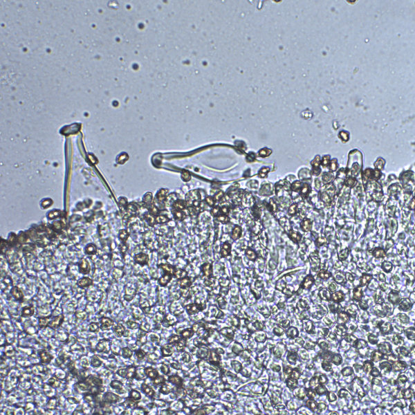
In order to look at some of the most interesting microscopic features of fungi, it is often useful to make a cross-section of the gill tissue. This makes it easier to observe some microscopic structures such as basidia and pleurocystidia.

There are many ways to make a gill section but the following method has proven the very useful.

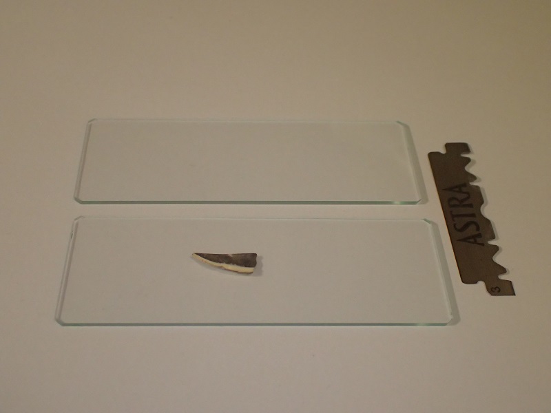
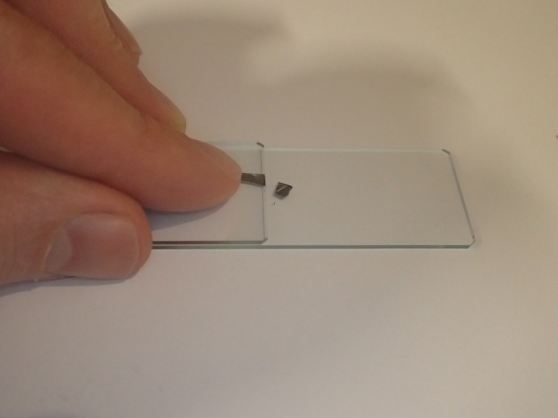
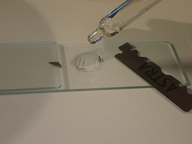
Essential items: 2 microscope slides, cover slips and a razor blade. A mushroom (from a shop is fine).

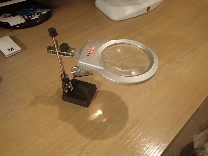
Recommended: Magnifier or stereomicroscope

Slides and gill
Slides and gill
An inexpensive magnifier

1. Cut a single gill from a mushroom using a razor blade and then cut off the excess cap material (if you don't do this then the section will not cut well) and the end sections.

Trimming
Trimming


Discard trimmed cap pieces leaving gill in place


2. Place another microscope slide on top and trim off a section of gill to leave a straight edge. For the very best results, use a BRAND NEW razor blade each time. You can label a used razor blade corner with a marker pen after use and use it for non-critical work.

Using the edge of the top slide like a ruler, carefully run the blade along the edge exerting downward pressure. Keep the razor vertical.


Discard excess gill
Discard excess gill

3. Now, using your left hand - slide the uppermost slide a fraction to the left so a very small piece of gill tissue is protruding. Using the top slide again like a ruler, make another vertical cut by dragging the vertical razor towards you. Repeat this several times. You may find the cut slices stick to the back of the razor. If this is the case, put a small drop of water on the right hand side of the lower slide and dip the blade in it. The sections should slip off straight into the water. Thin sections will fall right over in the water and lie flat.

A drop of water will make the thinner sections fall on their side
Steel needles or similar can be used to line the sections up and remove any that are too thick

4. Using forceps or a pin, gently lower a cover slip over the sample at an angle (to reduce trapped bubbles). Observe using a biological microscope.

Lay cover slip at an angle to reduce trapped air bubbles
Gill section laid on it's side.
Gill section laid on it's side. Observe fine structures with a biological microscope.
Mycena gill edge in Congo Red
Inocybe grammata pleurocystidia